Part 2
As more health care practitioners have understood the need to assess thyroid function based on what is going on at the cellular level, there has been an increasing demand for testing of reverse T3 (rT3), a hormone sometimes referred to as the “hibernation hormone.” However, there is also much confusion about how it fits into the picture of thyroid function, and controversy regarding whether or not there is a clinical utility for this test in patients suffering from thyroid imbalance symptoms.
The “hibernation hormone” that isn’t
Technically, the term “hibernation hormone” is inappropriate to describe reverse T3. Reverse T3 can be elevated in conditions associated with a reduction in the metabolic rate, notably starvation, extreme carbohydrate restriction, chronic heart failure, and the non-thyroidal illness syndrome (also called “euthyroid sick syndrome” or “low T3 syndrome”) seen in critical illness, very elderly patients, chronic stress, myocardial infarction, and chronic inflammatory states. In these cases, the rise in rT3 is a consequence, not a cause, of the alterations in intracellular thyroid hormone metabolism directed by the deiodinase enzymes, the relative activities of which are affected by the condition itself.
What is Reverse T3? Reverse T3 (3,3’,5’-triiodothyronine, rT3) is a biologically inactive metabolite of thyroxine (T4) formed by selective deiodination; the active thyroid hormone T3 is formed by removal of an iodine atom in the outer ring of T4, while rT3 is formed by removal of an iodine atom in the inner ring of T4. |
Relative amounts of each are determined by the activity of the respective deiodinase enzymes, which are regulated by hormonal and nutritional factors and physiological conditions.
Does rT3 really slow metabolism?
It is frequently claimed in articles published on the internet, but not in peer-reviewed papers, that rT3 blocks or “gets in the way of” the nuclear thyroid receptors. The nuclear thyroid receptors are the sites of action where the primary active thyroid hormone, T3, exerts its effects to drive cellular metabolism and maintain body temperature. There is no credible scientific evidence that rT3 enters the nucleus of the cell at all, and the bulk of the scientific literature states clearly that rT3 does not bind to, and has no known transcriptional activity at, the thyroid receptor. It is, however, known to have potent activity in the cytoplasm as an initiator of actin polymerization in astrocytes in the brain [7]. This is mediated in a non-genomic manner by its binding to a very specific thyroid receptor that exists only in the extranuclear compartment. Actin polymerization is important to cell structure and motility, and particularly important to normal brain development.
What do high levels of rT3 relative to T3 indicate?
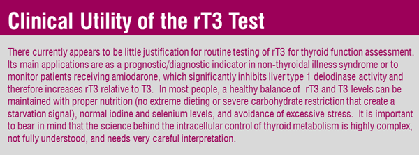
Any interpretation of the relative levels of T3 and rT3 must take into account all the factors that affect the activity of all three deiodinases. As explained in Part 1, reactivation of the D3 deiodinase and downregulation of the D1 deiodinase contribute to both increased rT3 formation and reduced rT3 clearance, while at the same time reducing T3 synthesis from T4, so conditions affecting the expression of D1 and D3 in this way have a profound impact on circulating levels of T3 and rT3 and the T3/rT3 ratio. The T3/rT3 ratio has therefore been found to have some value in assessing prognosis in severely ill [8] or very elderly patients [9]. There is currently no evidence for a clinical basis for the use of this ratio in routine thyroid function assessment.
Some observed elevations in serum rT3 in the presence of exogenous thyroxine treatment are difficult to interpret, and can depend on the assay used. T4 can cross-react in immunoassays for rT3, resulting in a false increase in rT3 when T4 is high; more reliable tests for rT3 are performed using liquid chromatography/tandem mass spectrometry (LC-MS/MS). A study in euthyroid subjects receiving thyroxine suggested that the observed rise in rT3 was a result of increased substrate availability for the peripheral inactivation of T4 [10]. And elderly patients may either have an adaptive response to reduce thyroid hyperactivity at the tissue level, or have some type of non-thyroidal illness with consequent reactivation of D3, in both cases resulting in increased T4 inactivation via conversion to rT3; it has therefore been suggested that older patients with hypothyroidism may require lower replacement doses of T4 [11].

Want to learn about the Deiodinases & Thyroid Hormone Bioavailability? Read Part I of this blog post.
Related Resources
References
